Microscopic Image Of Plant Cell Labelled : Immunogold Labelling Wikipedia : Open stax (rice university) and biology online.
byHal Innes-
0
Microscopic Image Of Plant Cell Labelled : Immunogold Labelling Wikipedia : Open stax (rice university) and biology online.. Going back to the original question, in plant cell preparations like this, what we see as the boundary around each cell is. In a plant cell the vacuole is filled with cellular juices, which dissolve various organic and inorganic substances. Affordable and search from millions of royalty free images, photos and vectors. Biology laboratory nature and science, plants with biochemistry structure on green background. Microscopic images of flesh cells of lyq loquat fruit.
Going back to the original question, in plant cell preparations like this, what we see as the boundary around each cell is. Introduction cells are the basic units of all living organisms. This work focuses on quantification analysis of plant cells in microscopic images the goal of this work is to find the dynamic interection between different structures in plant cells current work includes segmenting filamentous structures at. Flickr is almost certainly the best online photo bovine pulmonary artery endothelial cells cells are often fixed (and therefore dead) prior to labeling with fluorescent probes. If you see some purple pigment on your microscopic picture.
A Large Scale Optical Microscopy Image Dataset Of Potato Tuber For Deep Learning Based Plant Cell Assessment Scientific Data from media.springernature.com Flickr is almost certainly the best online photo bovine pulmonary artery endothelial cells cells are often fixed (and therefore dead) prior to labeling with fluorescent probes. The right and left images show different viewing angles. Find the perfect microscopic cell stock photos and editorial news pictures from getty images. In young cells, such vacuoles are picture 3. We say cells are microscopic because they can only be seen under a microscope. Fluorescent labeling of plastids simultaneously with other subcellular locations reveals dynamic interactions and mutant phenotypes. Download plant cell stock photos. The cell (plasma) membrane is about 7.5 nanometers (nm) thick, and the light microscope doesn't allow us to resolve , former professor of histology (microscopic anatomy).
In a plant cell the vacuole is filled with cellular juices, which dissolve various organic and inorganic substances.
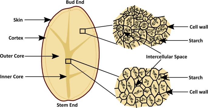
The right and left images show different viewing angles. Plants store starch grains within the cytoplasm. You can get your learners to provide labels for the vacuoles and. Download plant cell stock photos. An intricately illustrated 160 plus year old botanical print in black and white showing plant cells. A sclerenchyma cell with highly thickened wall; B single solid sclerenchyma cell; Fluorescent labeling of plastids simultaneously with other subcellular locations reveals dynamic interactions and mutant phenotypes. With proper selection of fluorophores for labeling the components of the plant cell, confocal microscopy can produce extremely informative images at. Vacuole can be reduced or increased depending on environmental conditions. Welcome to the inside of plant cells. In a plant cell the vacuole is filled with cellular juices, which dissolve various organic and inorganic substances. Microscopic plant cells (69 images).
Label the diagram and differences table:this is a great supplement for students to review/assess and strengthen their knowledge the unit of animal and plant cell unit. Nuclear magnetic resonance imaging of a single cell. Browse 10,546 microscopic cell stock photos and images available, or search for microscopic image or cancer cell to find more great. Mitochondria are sometimes described as cellular power plants because they produed most of the cells chemical energy in form of adenosine triphosphate(atp). (d) interface of the lipservice.
Magnification Bioninja from ib.bioninja.com.au Plant cell under the microscope view for education. See more ideas about microscopic photography, microscopic images, microscopic. Nuclear magnetic resonance imaging of a single cell. Fluorescent labeling of plastids simultaneously with other subcellular locations reveals dynamic interactions and mutant phenotypes. Label the diagram and differences table:this is a great supplement for students to review/assess and strengthen their knowledge the unit of animal and plant cell unit. Cell is a tiny structure and functional unit of a living organism containing various parts known as organelles. Affordable and search from millions of royalty free images, photos and vectors. Browse 10,546 microscopic cell stock photos and images available, or search for microscopic image or cancer cell to find more great.
Biology laboratory nature and science, plants with biochemistry structure on green background.
Plants store starch grains within the cytoplasm. Testing result of the proposed framework shows that it can achieve good performance in automatic cell detection and classification. Microscopic preparation, tissue plant, stomata. It is … read more. Huge collection, amazing choice, 100+ million high quality, affordable rf and rm images. Welcome to the inside of plant cells. See more ideas about microscopic photography, plant cell, microscopic images. If you see some purple pigment on your microscopic picture. Plant cell under the microscope view for education. Mitochondria are sometimes described as cellular power plants because they produed most of the cells chemical energy in form of adenosine triphosphate(atp). Going back to the original question, in plant cell preparations like this, what we see as the boundary around each cell is. Label the diagram and differences table:this is a great supplement for students to review/assess and strengthen their knowledge the unit of animal and plant cell unit. Browse 10,546 microscopic cell stock photos and images available, or search for microscopic image or cancer cell to find more great.
Plants store starch grains within the cytoplasm. Biology laboratory nature and science, plants with biochemistry structure on green background. (d) interface of the lipservice. See more ideas about microscopic, plant cell, microscopic photography. Label the diagram and differences table:this is a great supplement for students to review/assess and strengthen their knowledge the unit of animal and plant cell unit.
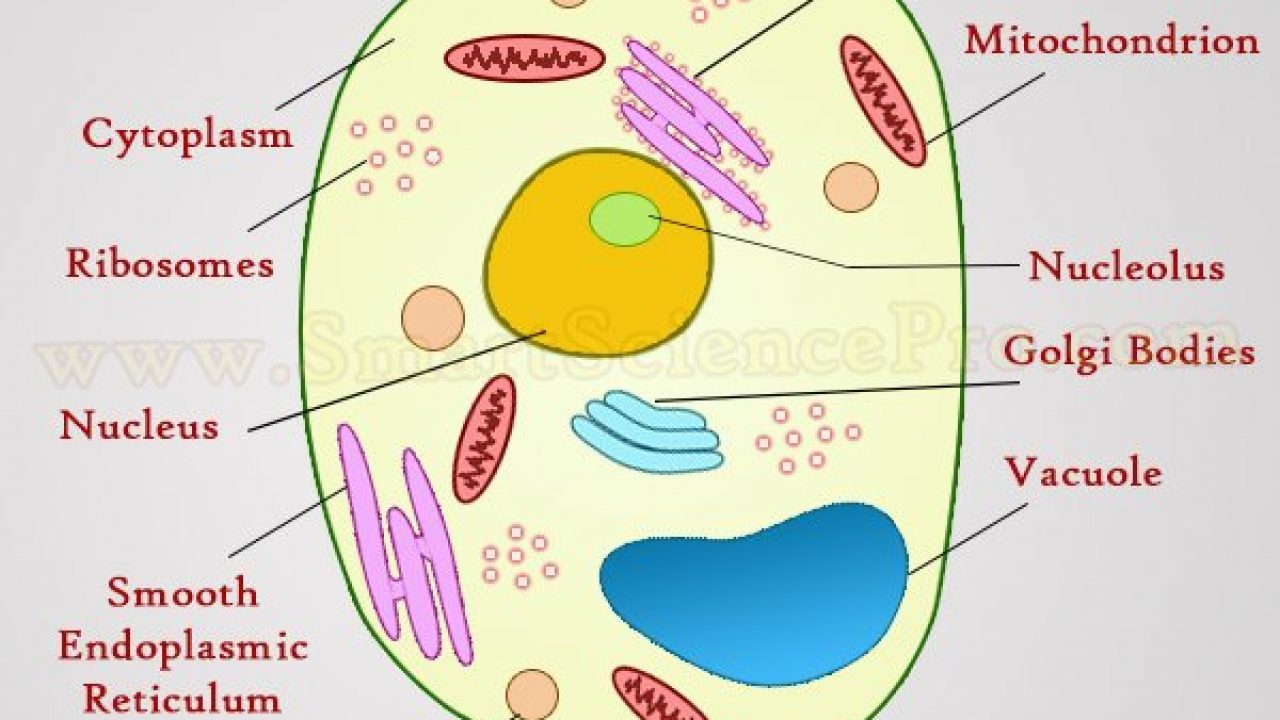
Structure Of Animal Cell And Plant Cell Under Microscope Diagrams from www.smartsciencepro.com Microscopic images of flesh cells of lyq loquat fruit. Mitochondria are sometimes described as cellular power plants because they produed most of the cells chemical energy in form of adenosine triphosphate(atp). Testing result of the proposed framework shows that it can achieve good performance in automatic cell detection and classification. Cells determine the organization of all plants and animals. Cell is a tiny structure and functional unit of a living organism containing various parts known as organelles. Plant cells contain many organelles such as ribosomes, the nucleus, the plasma membrane, the cell wall, mitochondria, and chloroplasts. Download plant cell stock photos. Plants store starch grains within the cytoplasm.
The cell (plasma) membrane is about 7.5 nanometers (nm) thick, and the light microscope doesn't allow us to resolve , former professor of histology (microscopic anatomy).
The right and left images show different viewing angles. Introduction cells are the basic units of all living organisms. Mitochondria are sometimes described as cellular power plants because they produed most of the cells chemical energy in form of adenosine triphosphate(atp). In this image of a plant cell, there are several vacuoles present, as is the case in many plant cells. Label the diagram and differences table:this is a great supplement for students to review/assess and strengthen their knowledge the unit of animal and plant cell unit. Cells determine the organization of all plants and animals. This work focuses on quantification analysis of plant cells in microscopic images the goal of this work is to find the dynamic interection between different structures in plant cells current work includes segmenting filamentous structures at. Microscopic plant cells (69 images) microscopic plant cells. Labeled plant cell with diagrams. Browse 10,546 microscopic cell stock photos and images available, or search for microscopic image or cancer cell to find more great. Perfect for framing or perhaps a science lesson! Testing result of the proposed framework shows that it can achieve good performance in automatic cell detection and classification. See more ideas about microscopic photography, plant cell, microscopic images.